Fetal Echocardiography Advanced
3-D and 4-D Ultrasound
What Is Fetal Echocardiography?
Fetal echocardiography is a test similar to an ultrasound. This exam allows your doctor to better see the structure and function of your unborn child’s heart. It’s typically done in the second trimester, between weeks 18 to 24.
The exam uses sound waves that "echo" off of the structures of the fetus’ heart. A machine analyzes these sound waves and creates a picture, or echocardiogram, of their heart’s interior. This image provides information on how your baby’s heart has formed and whether it’s working properly.
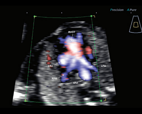
It also allows your doctor to see the blood flow through their heart. This in-depth look allows your doctor to find any defects or abnormalities in the baby’s blood flow or heartbeat.
Fetal echocardiography is the primary modality for defining and evaluating fetal cardiac status and requires detailed analysis of the cardiac anatomy from numerous views and Doppler interrogation of the intracardiac structures, great vessels, and umbilical artery. Referrals for fetal echocardiography are determined by fetal, maternal, or familial risk factors; however, approximately 50% of neonates diagnosed with a congenital cardiac defect have no risk factor, and most have undergone an obstetrical ultrasound during the pregnancy that did not detect a cardiac defect. Advances in transducer technology have resulted in the development of small high-frequency transvaginal probes that allow fetal cardiac interrogation earlier during gestation. On the horizon is 3-dimensional fetal echocardiography, which provides rapid image acquisition and tremendous computer image reconstruction ability. At present, the computer image data analysis process is lengthy, and several technical limitations must be overcome before 3-dimensional fetal echocardiography becomes the primary modality of fetal cardiac imaging. New Doppler Tissue Imaging using color Doppler energy mapping allows more precise anatomic definition of the fetal endocardium, facilitating diagnosis of small ventricular septal defects. These new advances, along with improved image resolution, provide obstetricians and pediatric cardiologists with more tools and techniques for earlier and more precise detection of fetuses with cardiac defects.